
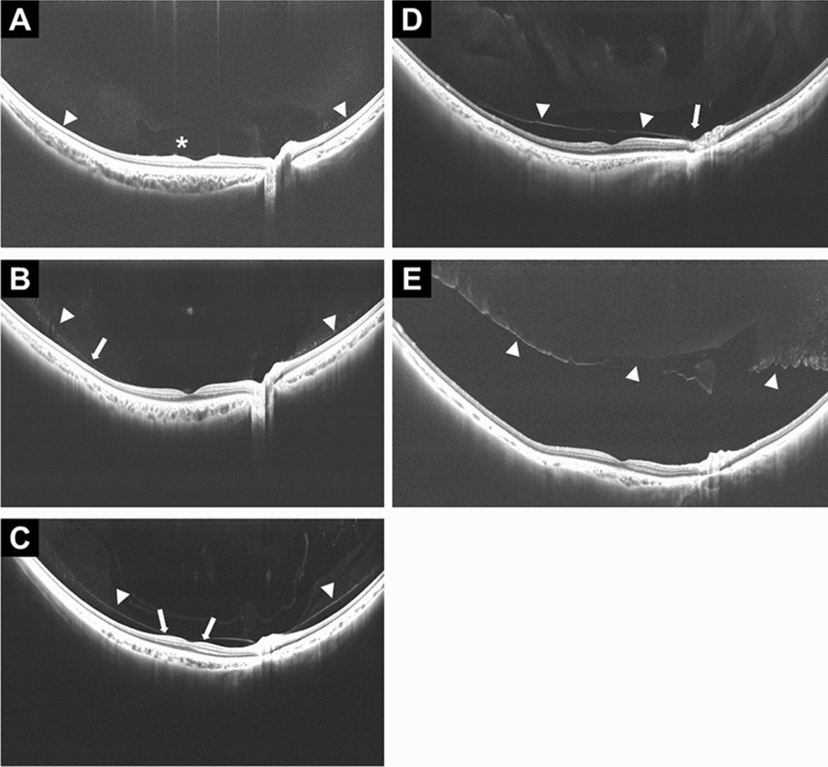
Figure 1. Stages of posterior vitreous detachment on widefield swept-source OCT, from vitreous attached to fully separated. Image: Chiku Y, et al. Sci Rep 2021;11:19754, Fig. 3, CC BY 4.0. Source
What is it?
The inside of the eye is filled with a clear gel called the vitreous. With age, this gel becomes more watery and pulls away from the retina, the light-sensing layer at the back of the eye. This is called a posterior vitreous detachment, or PVD. It is a normal part of aging and happens to most people eventually. It can cause new floaters (spots, cobwebs or a ring drifting in your vision) and brief flashes of light, especially in dim light.
What do I do about it?
A PVD itself does not need treatment. Floaters usually fade or become less noticeable over a few weeks to months. The main risk is that the gel tugs hard enough to tear the retina, which happens in about 1 in 7 people with new symptoms. That is why I examine the whole retina with your pupils dilated, and why I ask you to come back in a few weeks for a second look, since a tear can show up later.
When should I call?
Call my office the same day at 408-540-3559 if you notice:
- A sudden shower of new floaters
- New or more frequent flashes of light
- A shadow or curtain coming over part of your vision
- Sudden blurry or lost vision
This page is for general education and does not replace an exam. Call Cai Retina at 408-540-3559 with questions.